Bronzite HS9.3B — NIR2,126 points · 4000–12500 cm⁻¹
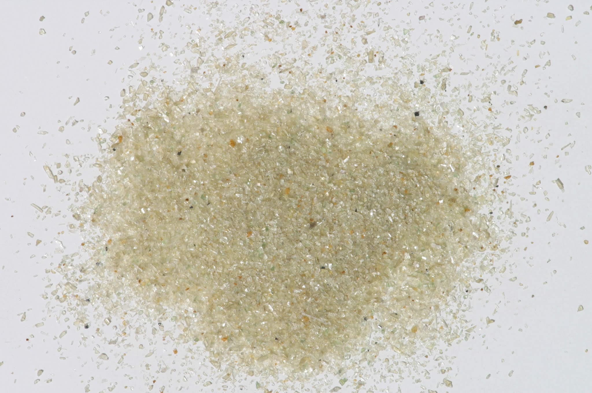
Sample photograph — USGS Spectral Library v7
Spectrum Details
- Modality
- NIR
- Category
- mineral
- Material Type
- Inosilicate
- Sample ID
- HS9.1B, HS9.2B, HS9.3B, HS9.4B
- Collection Locality
- Jackson Co., North Carolina
- Spectral Purity
- 1b2_3_4_ # Bronzite HS9.1B # 1= 0.2-3, 2= 1.5-6, 3= 6-25, 4= 20-150 microns SPECTRAL_PURITY: 1c2_3_4_ # Bronzite HS9.2B # 1= 0.2-3, 2= 1.5-6, 3= 6-25, 4= 20-150 microns SPECTRAL_PURITY: 1c2c3c4c # Bronzite HS9.3B # 1= 0.2-3, 2= 1.5-6, 3= 6-25, 4= 20-150 microns SPECTRAL_PURITY: 1c2_3_4_ # Bronzite HS9.4B # 1= 0.2-3, 2= 1.5-6, 3= 6-25, 4= 20-150 microns
- Composition / XRD
- None # XRF, EPMA, ICP(Trace), WChem
- Sample Description
- Spectrum displays double ferrous iron band typical of iron-rich orthopyroxenes. Original spectrum published in: Hunt, G.R., J.W. Salisbury, 1970, Visible and near-infrared spectra of minerals and rocks: I. Silicate minerals. Modern Geology, v. 1, p. 283-300. "S-17E. Pyroxene, variety Bronzite. Jackson CO, N. Carolina (9B). This sample also displays the double ferrous ion band in the near-infrared typical of iron-rich orthopyroxenes. The slight band in the visible at 0.5µm is also a ferrous ion band." Sieve interval 74 - 250µm. Weak absorptions at 1.4 and 2.3 µm indicate trace alteration/contamination (probably tremolite). Roger N. Clark Grain size fractions are indicated by the extension after the sample number: .1B = IMAGE_OF_SAMPLE:
- XRD Analysis
- 40 kV - 30 mA, 6.5-9.5 keV File: brnzt9.out, -.mdi; brnzt9b.out, -.mdi (at low angles) (Both patterns obtained from smear mounts on quartz plates) References: Huebner's reference patterns Found: orthopyroxene, minor talc, trace chlorite (?), many un indexed moderately to very weak reflections (6.35, 5.25, 3.90, 3.58, 3.27, 3.23, 2.775, 2.255, 1.888, 1.643, and 1.580 Angstroms) Comment: Sharp, strong pattern of orthopyroxene. Talc based on four basal reflections. Needs more work.
- X Units
- cm⁻¹
- Y Units
- Absorbance
- Data Points
- 2,126
Download
Attribution
USGS Spectral Library Version 7, U.S. Geological Survey
License: Public Domain
DOI: 10.3133/ds1035Analyze in Workbench
Open this spectrum in the SpectralBench workbench for advanced analysis, peak detection, and comparison.
Open Workbench