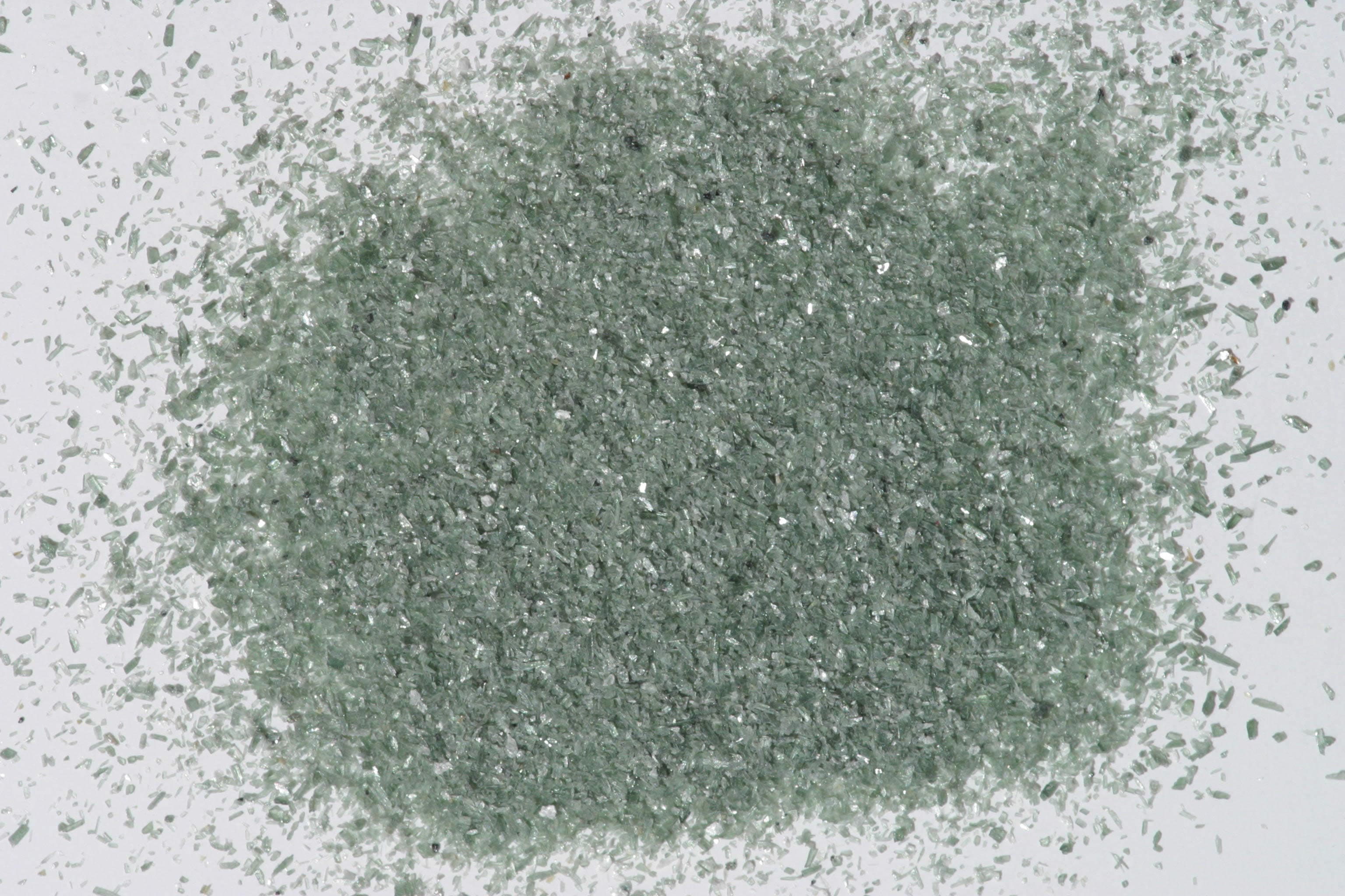
Chlorite — FTIR1,801 points · 400–4000 cm⁻¹
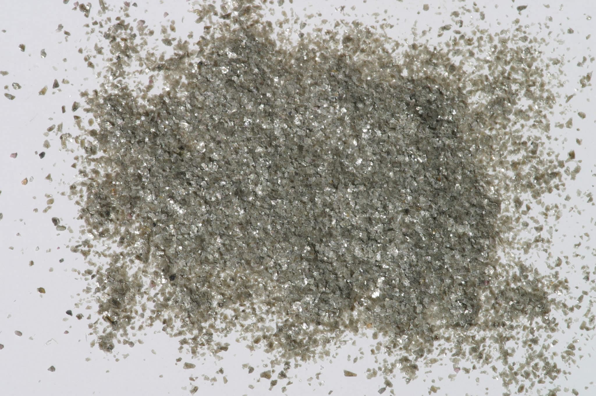
Sample photograph — USGS Spectral Library v7
Spectrum Details
- Modality
- FTIR
- Category
- mineral
- Material Type
- Phyllosilicate
- Sample ID
- HS179.1B, HS179.2B, HS179.3B, HS179.4B, HS179.6
- Collection Locality
- Colorado
- Spectral Purity
- 1b2_3_4_ # Chlorite HS179.1B # 1= 0.2-3, 2= 1.5-6, 3= 6-25, 4= 20-150 microns SPECTRAL_PURITY: 1b2_3_4_ # Chlorite HS179.2B # 1= 0.2-3, 2= 1.5-6, 3= 6-25, 4= 20-150 microns SPECTRAL_PURITY: 1b2b3b4u # Chlorite HS179.3B # 1= 0.2-3, 2= 1.5-6, 3= 6-25, 4= 20-150 microns SPECTRAL_PURITY: 1b2_3_4_ # Chlorite HS179.4B # 1= 0.2-3, 2= 1.5-6, 3= 6-25, 4= 20-150 microns SPECTRAL_PURITY: 1b2_3_4_ # Chlorite HS179.6 # 1= 0.2-3, 2= 1.5-6, 3= 6-25, 4= 20-150 microns
- Composition / XRD
- None # XRF, EPMA, ICP(Trace), WChem
- Sample Description
- A spectrum for this sample was published in: Hunt, G.R., J.W. Salisbury, 1970, Visible and near-infrared spectra of minerals and rocks: I. Silicate minerals. Modern Geology, v. 1, p. 283-300. With the note: "This specimen appears to be quite pure. The spectrum displays three broad bands between 0.7 and 1.1µm. The bands at 0.7µm (barely discernible in this spectrum but very marked in the following sample (HS197) and at 0.9µm are due to Fe3+ in six fold coordination. The band near 1.1µm is due to the ferrous ion in six fold coordination. The sharp features at 1.4µm and between 2.0 and 2.6 µm are hydroxyl bands as in previous samples." For additional information on spectral features of chlorites see: King, T.V.V. and R.N. Clark, 1989, Spectral Characteristics of Chlorites and Mg-Serpentines Using high-Resolution Reflectance Spectroscopy. Jour. Geophys. Res., 13,997-14,008. Grain size fractions are indicated by the extension after the sample number: .1B = IMAGE_OF_SAMPLE:
- XRD Analysis
- 40 kV - 30 mA, 7.0-9.5 keV File: chlor179.mdi, *.out References: Bailey (198-, MSA vol.13, p.557); Huebner's reference patterns Found: Tri-octahedral mica, trace 7 or 14 Angstrom phase, and an un indexed weak reflection at 3.71 Angstroms Comment: The mica could be indexed as a 1M phlogopite; the pattern is very similar to biotite MBLM. Reflections at 7.2 and 3.54 Angstroms are consistent with a chlorite or a kaolinite mineral. No 14 Angstroms reflection was observed.
- X Units
- cm⁻¹
- Y Units
- Absorbance
- Data Points
- 1,801
Download
Attribution
USGS Spectral Library Version 7, U.S. Geological Survey
License: Public Domain
DOI: 10.3133/ds1035Analyze in Workbench
Open this spectrum in the SpectralBench workbench for advanced analysis, peak detection, and comparison.
Open Workbench