Ferrihydrite — NIR2,126 points · 4000–12500 cm⁻¹
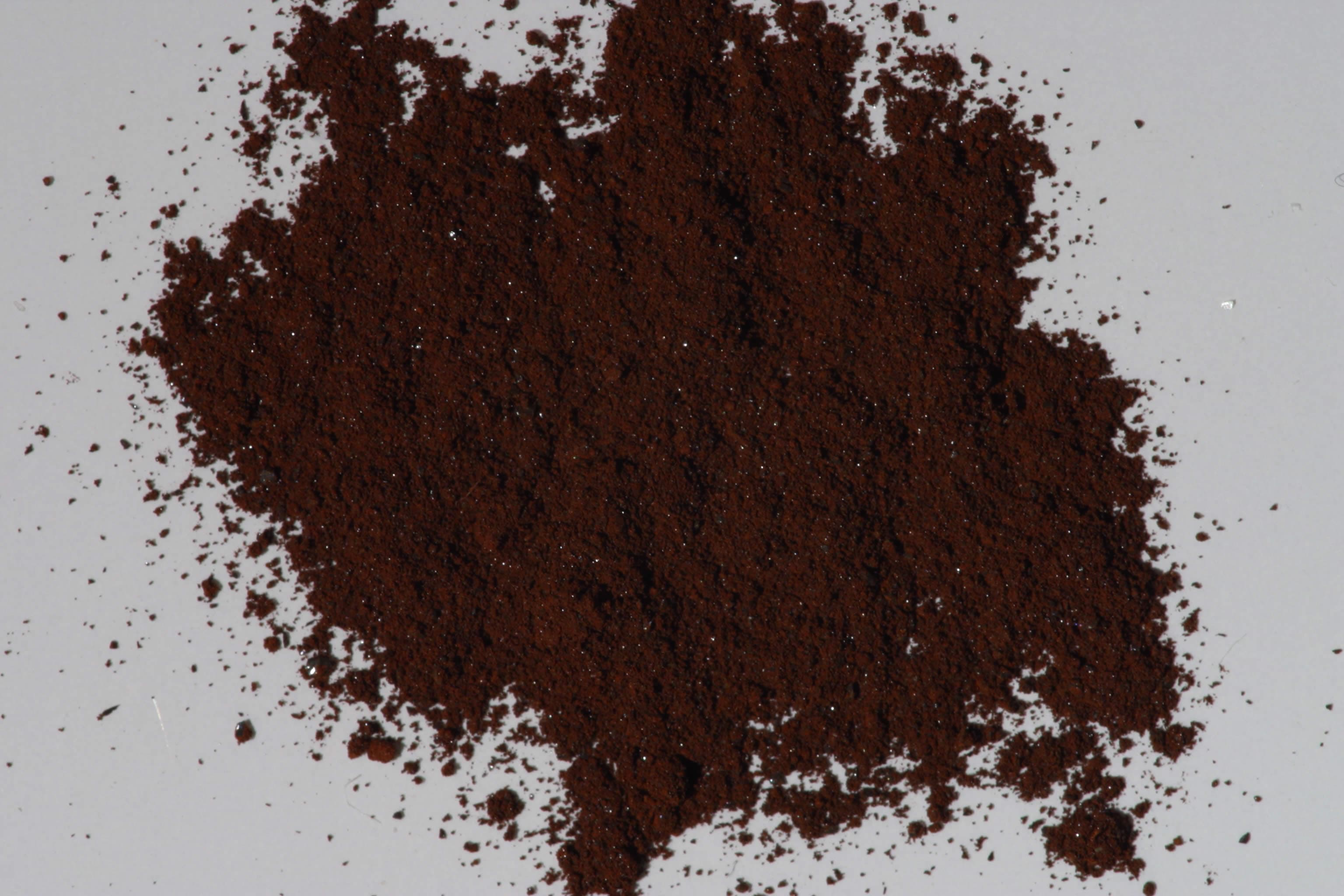
Sample photograph — USGS Spectral Library v7
Spectrum Details
- Modality
- NIR
- Category
- mineral
- Material Type
- Oxide
- Sample ID
- GDS75 (F6)
- Collection Locality
- Synthetic
- Spectral Purity
- 1b2_3_4_ # 1= 0.2-3, 2= 1.5-6, 3= 6-25, 4= 20-150 microns
- Composition / XRD
- # XRF, EPMA, ICP(Trace), WChem
- Sample Description
- This sample is one of a series of ferrihydrite samples made by Dave Sherman for the following paper: Sherman, D.M., R.G. Burns, and V.M. Burns, 1982, Spectral characteristics of the iron oxides with application to the Martian bright region mineralogy. Journal of Geophysical Research, v. 87, n. B12, pp. 10169- 10180. (F6 is Dave Sherman's number) IMAGE_OF_SAMPLE:
- XRD Analysis
- the sample is x-ray amorphous - G. Swayze and Ken Esposito. 40 kV - 30mA, 7.3-9.5 kev File: fehyd75.mdi (smear mount on quartz plate) References: JCPDS #29-712 Found: Amorphous to CuKalpha X-radiation Comment: Pattern has a high background but no significant reflections. A weak, very broad maximum is centered around 2.6 Angstroms (Fe-O bond?); an even weaker broad maximum centered about 1.50 Angstroms could be a sample response or an instrumental effect.
- X Units
- cm⁻¹
- Y Units
- Absorbance
- Data Points
- 2,126
Download
Attribution
USGS Spectral Library Version 7, U.S. Geological Survey
License: Public Domain
DOI: 10.3133/ds1035Analyze in Workbench
Open this spectrum in the SpectralBench workbench for advanced analysis, peak detection, and comparison.
Open Workbench