Gibbsite — FTIR1,801 points · 400–4000 cm⁻¹
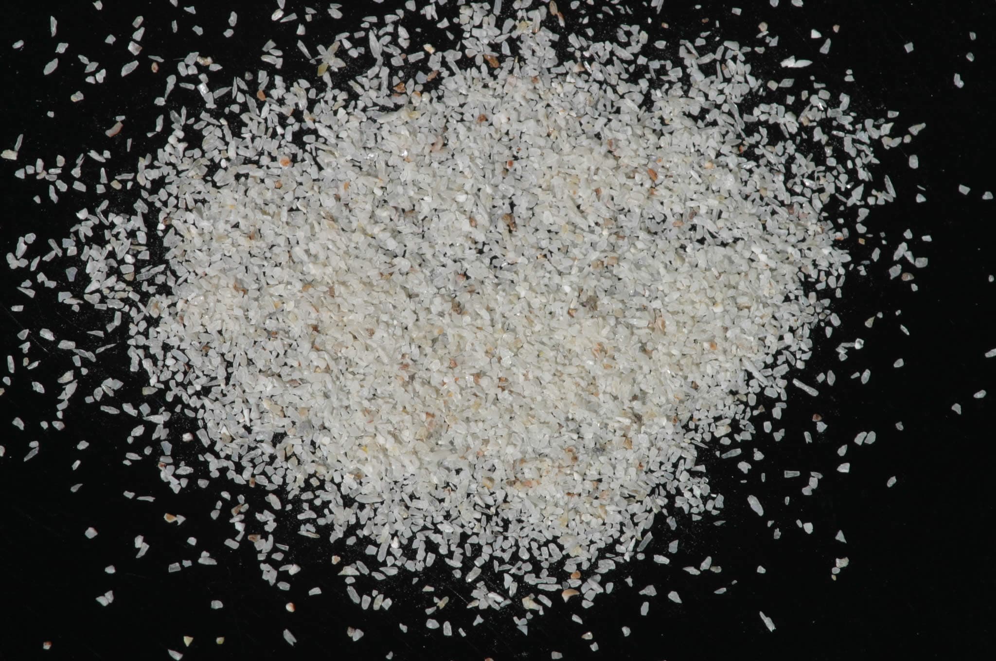
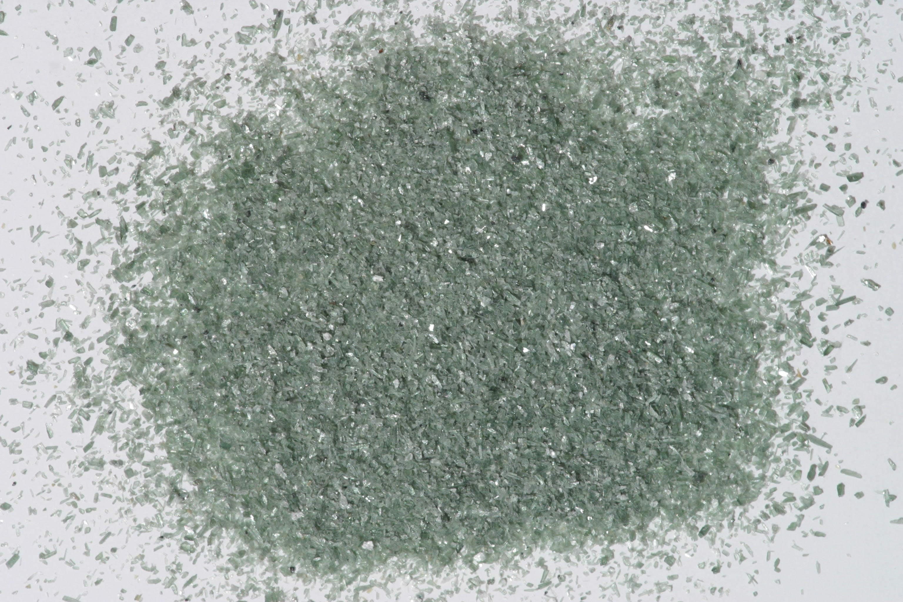
Sample photograph — USGS Spectral Library v7
Spectrum Details
- Modality
- FTIR
- Category
- mineral
- Material Type
- Hydroxide
- Sample ID
- HS423.1B, HS423.2B, HS423.3B, HS423.4B, HS423.6
- Collection Locality
- Brazil
- Spectral Purity
- 1b2_3_4_ # Gibbsite HS423.1B # 1= 0.2-3, 2= 1.5-6, 3= 6-25, 4= 20-150 microns SPECTRAL_PURITY: 1b2_3_4_ # Gibbsite HS423.2B # 1= 0.2-3, 2= 1.5-6, 3= 6-25, 4= 20-150 microns SPECTRAL_PURITY: 1b2b3b4b # Gibbsite HS423.3B # 1= 0.2-3, 2= 1.5-6, 3= 6-25, 4= 20-150 microns SPECTRAL_PURITY: 1b2_3_4_ # Gibbsite HS423.4B # 1= 0.2-3, 2= 1.5-6, 3= 6-25, 4= 20-150 microns SPECTRAL_PURITY: 1b2_3_4_ # Gibbsite HS423.6 # 1= 0.2-3, 2= 1.5-6, 3= 6-25, 4= 20-150 microns
- Composition / XRD
- None # XRF, EPMA, ICP(Trace), WChem
- Sample Description
- Trimorphous with Bayerite and Nordstrandite. Original spectrum published in: Hunt, G.R., J.W. Salisbury, and C.J. Lenhoff, 1971, Visible and near-infrared spectra of minerals and rocks: III. Oxides and hydroxides. Modern Geology, v. 2, p. 195-205. With the comment:"This particular sample is a cryptocrystalline stalactitic aggregate, composed of concentric layers of gibbsite of variable purity. Some layers are slightly contaminated with iron, which produces the gentle fall-off in reflectivity of the powdered samples toward the blue region of the visible. In the near-infrared, all the spectral features are due to the vibrations of the hydroxyl ion. In the mid-infrared, well defined fundamental Al-O-H bending modes occur at 10.34 and 9.8µm, and a series of fundamental stretching modes occur at 2.975, 2.917, 2.842, and 2.765µm. The second overtone and combinations of the stretching modes produce the weak but relatively sharp features which appear near 1.0µm in our spectra, the first overtone and combinations of the stretching mode produce the numerous relatively sharp features centered at 1.45µm, and the combinations of the stretching and bending modes produce the features near 2.3µm. Some free water is indicated by the bands near 1.9µm." Grain size fractions are indicated by the extension after the sample number: .1B = IMAGE_OF_SAMPLE:
- XRD Analysis
- Pure gibbsite Konnert, Judith and Marta Flohr, 1992, unpublished data, USGS Reston, VA.
- X Units
- cm⁻¹
- Y Units
- Absorbance
- Data Points
- 1,801
Download
Attribution
USGS Spectral Library Version 7, U.S. Geological Survey
License: Public Domain
DOI: 10.3133/ds1035Analyze in Workbench
Open this spectrum in the SpectralBench workbench for advanced analysis, peak detection, and comparison.
Open Workbench