Knebelite — FTIR1,801 points · 400–4000 cm⁻¹
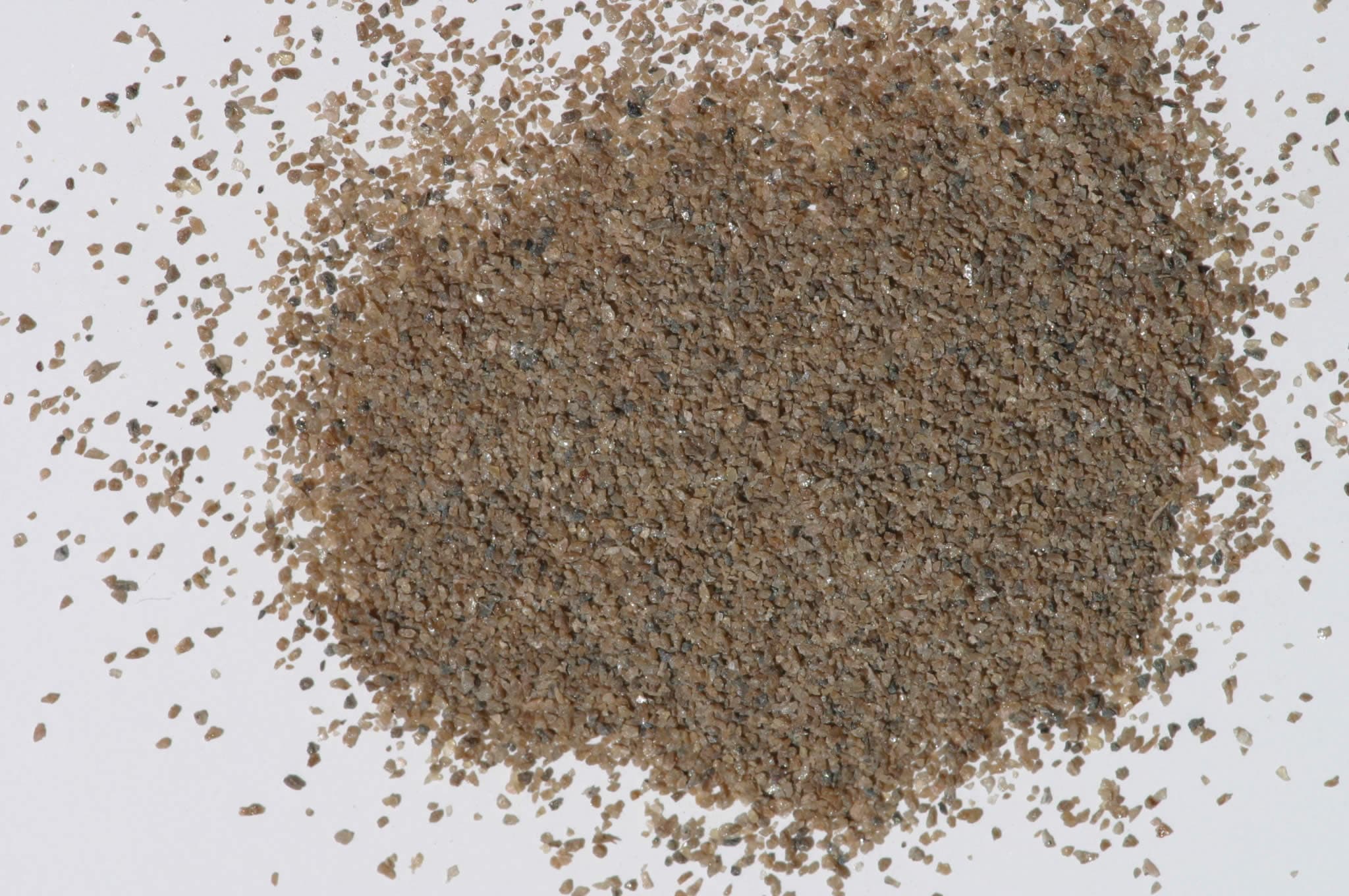
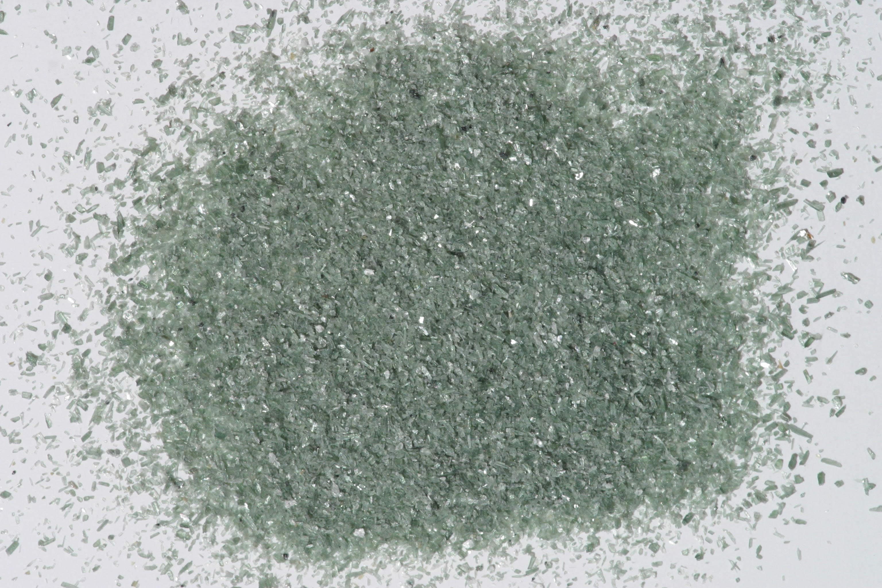
Sample photograph — USGS Spectral Library v7
Spectrum Details
- Modality
- FTIR
- Category
- mineral
- Material Type
- Nesosilicate
- Sample ID
- HS419.1B, HS419.2B, HS419.3B, HS419.4B, HS419.6
- Collection Locality
- Japan
- Spectral Purity
- 1b2_3_4_ # HS419.1B # 1= 0.2-3, 2= 1.5-6, 3= 6-25, 4= 20-150 microns SPECTRAL_PURITY: 1b2_3_4_ # HS419.2B # 1= 0.2-3, 2= 1.5-6, 3= 6-25, 4= 20-150 microns SPECTRAL_PURITY: 1c2c3b4b # HS419.3B # 1= 0.2-3, 2= 1.5-6, 3= 6-25, 4= 20-150 microns SPECTRAL_PURITY: 1c2_3_4_ # HS419.4B # 1= 0.2-3, 2= 1.5-6, 3= 6-25, 4= 20-150 microns SPECTRAL_PURITY: 1c2_3_4_ # HS419.6 # 1= 0.2-3, 2= 1.5-6, 3= 6-25, 4= 20-150 microns
- Composition / XRD
- None # XRF, EPMA, ICP(Trace), WChem COMPOSITION_TRACE:
- Sample Description
- Forms series with Fayalite. "N-5 Tephroite 419B--Japan: Ostensibly a manganese silicate, Mn2[SiO4], tephroite typically contains up to 10 percent iron. This sample is also contaminated with magnetite, which lowers its overall reflectivity. The manganese ion produces distinctive bands at 0.37, 0.415, 0.45, and 0.55 µ, which are subdued by the presence of the opaque magnetite, but are faintly discernible in the IV size range. The broad band near 1.0 µ, which continues out to near 1.7 µ, is due to the ferrous ion in substitution for Mn^2+; (see rhodochrosite, Part II, p. 28, spectrum C-6 for typical manganese-iron, p. 28, spectrum C-6 for typical manganese-iron spectra). The combination of the manganese and iron absorptions results in a well-defined reflectivity maximum near 0.73 µ." Hunt, G.R., J.W. Salisbury, and C.J. Lenhoff, 1973, Visible and near-infrared spectra of minerals and rocks: VI. Additional silicates. Modern Geology, v. 4, p. 85-106. Grain size fractions are indicated by the extension after the sample number: .1B = IMAGE_OF_SAMPLE:
- XRD Analysis
- 40 kV - 30 mA, 7.3-9.5 keV File: teph419.mdi (smear on quartz plate) References: PDF2 #12-0220, 35-0748; Huebner's reference patterns Found: Knebelite Sought but not found: alleghanyite, manganbabingdonite, nambulite, sonolite, pyroxmangite, rhodonite, santaclaraite, manganohumite, leucophenacite, hausmannite, marsturite, johannsenite Comments: Weak pattern with moderately sharp peaks but poor resolution of alpha1-alpha2 components. Shoulders and broadening of peak tips suggest compositional zoning. Moderately high background is consistent with Mn X-ray fluorescence. Twenty five reflections were identified as tephroite; 19 were refined resulting in cell dimensions a=6.195(1), b=10.600(2), and c=4.868(1) Angstroms. These dimensions are most consistent with ferroan or slightly mangesian tephroite. Subsequent profile-based search-match returned manganoan fayalite (12-0220). Additional reflections occur at 3.15, 3.12, 2.96, 2.92, 2.28, 2.12, and 1.613 Angstroms. No single mineral (or simple combinations of minerals) explains these seven reflections. Samples HS419.3B and HS419.4B have carbonate contamination but samples HS419.1B and HS419.2B do not have detectable carbonate contamination. G. Swayze.
- X Units
- cm⁻¹
- Y Units
- Absorbance
- Data Points
- 1,801
Download
Attribution
USGS Spectral Library Version 7, U.S. Geological Survey
License: Public Domain
DOI: 10.3133/ds1035Analyze in Workbench
Open this spectrum in the SpectralBench workbench for advanced analysis, peak detection, and comparison.
Open Workbench