Lepidolite — UV-Vis601 points · 200–800 nm
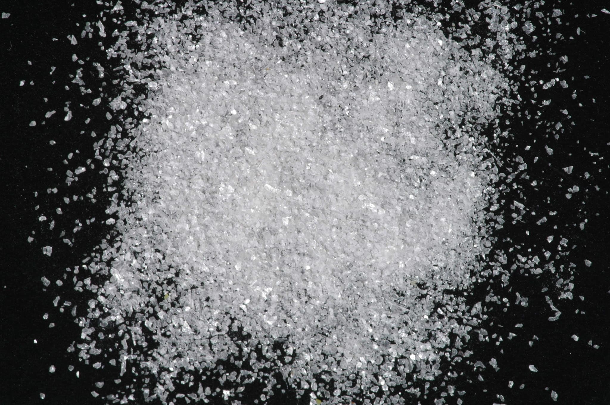
Sample photograph — USGS Spectral Library v7
Spectrum Details
- Modality
- UV-Vis
- Category
- mineral
- Material Type
- Phyllosilicate
- Sample ID
- HS167.1B, HS167.2B, HS167.3B, HS167.4B, HS167.6
- Collection Locality
- Keystone, S. Dakota
- Spectral Purity
- 1b2_3_4_ # HS167.1B # 1= 0.2-3, 2= 1.5-6, 3= 6-25, 4= 20-150 microns SPECTRAL_PURITY: 1b2_3_4_ # HS167.2B # 1= 0.2-3, 2= 1.5-6, 3= 6-25, 4= 20-150 microns SPECTRAL_PURITY: 1b2c3c4c # HS167.3B # 1= 0.2-3, 2= 1.5-6, 3= 6-25, 4= 20-150 microns SPECTRAL_PURITY: 1b2_3_4_ # HS167.4B # 1= 0.2-3, 2= 1.5-6, 3= 6-25, 4= 20-150 microns SPECTRAL_PURITY: 1b2_3_4_ # HS167.6 # 1= 0.2-3, 2= 1.5-6, 3= 6-25, 4= 20-150 microns
- Composition / XRD
- None # XRF, EPMA, ICP(Trace), WChem COMPOSITION_TRACE:
- Sample Description
- Original Spectrum published in: Hunt, G.R., J.W. Salisbury, and C.J. Lenhoff, 1973, Visible and near-infrared spectra of minerals and rocks: VI. Additional silicates. Modern Geology, v. 4, p. 85-106. With the note:"In addition to the substitutions given in the general formula for lepidolite, considerable amounts of sodium, rubidium and cesium may substitute for potassium; and iron, manganese and magnesium may substitute for aluminum. It is the substituted ferric iron and manganese that give rise to the bands at 0.8µ, 0.55 µ, and 0.45µ, together with the drop off to the extreme blue. The band near 1.4µ is due to the overtone of the OH stretching fundamental, and the very weak band at 1.9µ indicates that a small amount of included water is present. The bands near 2.2µ, 2.35µ, and 2.45µ are also due to combination bands involving the OH stretch and possibly the Al-O-H bending mode (at 2.2µ) as discussed above, and the very weak bands at 1.28µ, 1.33µ, 2.03µ and 2.14µ are OH combined with lattice modes." Grain size fractions are indicated by the extension after the sample number: .1B = IMAGE_OF_SAMPLE:
- XRD Analysis
- 40 kV - 30 mA, 6.5-9.5 keV File: lpd1673B.mdi (thick smear on quartz plate) References: Huebner's reference patterns; JCPDS patterns for micas; Deer, Howie, and Zussman (1962); Bailey (1984a,b); PDF2 #33-1161, 42-1399, 41-1480, 15-0062, 09-0466 Found: Quartz, a lepidolite-group mica, minor albite Comment: The pattern is dominated by strong, sharp quartz reflections. Mica reflections are sharp but not sufficiently intense to show clear separation of alpha1-alpha2 components, even at high 2 theta. The mica phase was compared to Huebner's reference patterns and the JCPDS mica indexes. Both 2M1 and 2M2 lepidolites and muscovites yield good matches. Search-match based on eight strong lines returns quartz, zinnwaldite, and calcian albite; little of the intensity of a reflection at 4.96 Angstroms was explained by zinnwaldite (002). Profile based search-match returned quartz, ordered albite, zinnwaldite, lepidolite-12O as likely hits. The reflection at 4.96 Angstroms is explained by lepidolite superlattice reflection (00.12); zinnwaldite contributes little to the interpretation of the pattern. Examination of the original sample shows a distinctive lavender-violet color implying that a member of the lepidolite mica group is present.
- X Units
- nm
- Y Units
- Cross-section
- Data Points
- 601
Download
Attribution
USGS Spectral Library Version 7, U.S. Geological Survey
License: Public Domain
DOI: 10.3133/ds1035Analyze in Workbench
Open this spectrum in the SpectralBench workbench for advanced analysis, peak detection, and comparison.
Open Workbench