Psilomelane — NIR2,126 points · 4000–12500 cm⁻¹
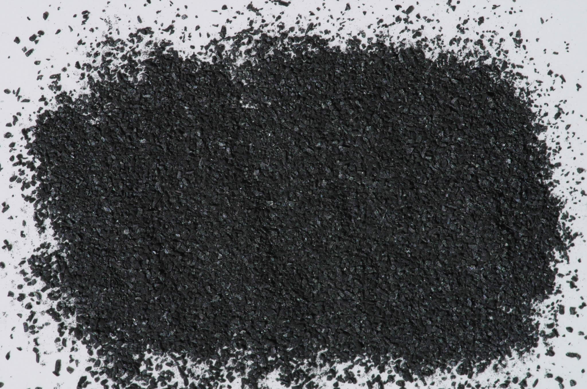
Sample photograph — USGS Spectral Library v7
Spectrum Details
- Modality
- NIR
- Category
- mineral
- Material Type
- Hydroxide (Mineral mixture)
- Sample ID
- HS139.1B, HS139.2B, HS139.3B, HS139.4B
- Collection Locality
- Magdalena, New Mexico
- Spectral Purity
- 1b2_3_4_ # HS139.1B # 1= 0.2-3, 2= 1.5-6, 3= 6-25, 4= 20-150 microns SPECTRAL_PURITY: 1b2_3_4_ # HS139.2B # 1= 0.2-3, 2= 1.5-6, 3= 6-25, 4= 20-150 microns SPECTRAL_PURITY: 1b2b3b4_ # HS139.3B # 1= 0.2-3, 2= 1.5-6, 3= 6-25, 4= 20-150 microns SPECTRAL_PURITY: 1b2_3_4_ # HS139.4B # 1= 0.2-3, 2= 1.5-6, 3= 6-25, 4= 20-150 microns
- Composition / XRD
- None # XRF, EPMA, ICP(Trace), WChem COMPOSITION_TRACE:
- Sample Description
- "0-13. Psilomelane. Magdalena, N.M. (139B). "Psilomelane" is generally used as a field term for any poorly characterized massive ore of manganese. Much of the material originally cleared as psilomelane consists of mixtures of several different minerals, usually with pyrolusite (Palache and others, 1944). Psilomelane usually is of supergene origin, occurring typically as a weathering product of manganiferous carbonates or silicates. This particular sample has a black streak, and yields very little water (0.09%) when heated. Consequently, we conclude that it is composed in large part of pyrolusite. Its spectrum is opaque and spectrally featureless, due to the conduction band of MnO2 extending throughout this spectral range." Hunt, G.R., J.W. Salisbury, and C.J. Lenhoff, 1971, Visible and near-infrared spectra of minerals and rocks: III. Oxides and hydroxides. Modern Geology, v. 2, p. 195-205. "Many of the hard botryoidal masses formerly called psilomelane are now known to be a mixture of several manganese oxides of which romanechite is a major constituent. Some of the other minerals commonly present in the mixture are cryptomelane, KMn8,O16; manjiroite, (Na,K)Mn8O16^.nH2O; and todorokite (Mn,Ca,Mg) Mn3O7^.H2O." Klein, C. and Hurlbut, C.S., Manual of Mineralogy 20th Edition, pp 317-318, 1985. Grain size fractions are indicated by the extension after the sample number: .1B = IMAGE_OF_SAMPLE:
- XRD Analysis
- 40 kV - 30 mA, 7.0-9.5 keV File: psilm139.mdi (smear on quartz plate) References: PDF2 #14-0627 Found: Romanechite Comments: All peaks are weak, most peaks broad, and the alpha1-alpha2 components are not resolved. The pattern has high background, probably due to Mn or Fe X-ray fluorescence. Within the range covered by the PDF2 data, all observed reflections were indexable as romanechite. There are some differences in intensity, however. The PDF2 lists additional reflections which were not observed in our poor pattern. Romanechite (Major), Akhtenskite(Minor)..Sutley April, 2005
- X Units
- cm⁻¹
- Y Units
- Absorbance
- Data Points
- 2,126
Download
Attribution
USGS Spectral Library Version 7, U.S. Geological Survey
License: Public Domain
DOI: 10.3133/ds1035Analyze in Workbench
Open this spectrum in the SpectralBench workbench for advanced analysis, peak detection, and comparison.
Open Workbench