Pyroxmangite — NIR2,126 points · 4000–12500 cm⁻¹
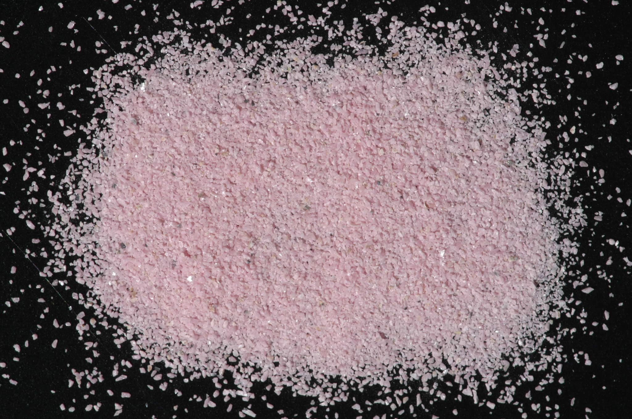
Sample photograph — USGS Spectral Library v7
Spectrum Details
- Modality
- NIR
- Category
- mineral
- Material Type
- Inosilicate
- Sample ID
- HS325.3B
- Collection Locality
- Colorado
- Spectral Purity
- 1b2b3b4_ # HS325.3B # 1= 0.2-3, 2= 1.5-6, 3= 6-25, 4= 20-150 microns
- Composition / XRD
- None # XRF, EPMA, ICP(Trace), WChem COMPOSITION_TRACE:
- Sample Description
- This sample contains very small amounts of magnetite, pyrolusite and calcite. The visible spectrum is dominated by features typical of Mn^2+ transitions, displaying bands at 0.35 µ, 0.37 µ, 0.42 µ, and 0.55 µ, resulting in the characteristic pink color of this mineral. The strong broad band near 1.04 µ and 1.9 µ features are typical of molecular water, probably in fluid inclusions. Hunt and others (1973) mis-identified this sample as rhodonite. XRD analysis indicates it is pyroxmangite, a polymorph of rhodonite. Hunt, G.R., J.W. Salisbury, and C.J. Lenhoff, 1973, Visible and near-infrared spectra of minerals and rocks: VI. Additional silicates. Modern Geology, v. 4, p. 85-106. IMAGE_OF_SAMPLE:
- XRD Analysis
- 40 kV - 30 mA, 7.3-9.5 keV File: rhod325.mdi References: Huebner's reference patterns; PDF2 #29-0985 Found: Pyroxmangite Comments: Peaks are symmetric but not strong; alpha1-alpha2 are not resolved. The pattern has a high background caused by X-ray fluorescence (Mn- or Fe-rich composition is probable). HS-325 is very similar to Huebner's reference patterns for synthetic pyroxmangite of MnSiO3 composition (runs Hy-67 and run-130) and unlike that of rhodonite (run49; see Heubner, 1986). Profile based search-match returns pyroxmangite (PDF2 #29-0985) There are weak, very sharp reflections at 7.2 and 1.912 Angstroms but no mineral in the PDF2 database has two strong reflections at these positions. Additional XRD analysis is consistent with pyroxmangite and not rhodonite. B. Benzel.
- X Units
- cm⁻¹
- Y Units
- Absorbance
- Data Points
- 2,126
Download
Attribution
USGS Spectral Library Version 7, U.S. Geological Survey
License: Public Domain
DOI: 10.3133/ds1035Analyze in Workbench
Open this spectrum in the SpectralBench workbench for advanced analysis, peak detection, and comparison.
Open Workbench