Rutile — NIR2,126 points · 4000–12500 cm⁻¹
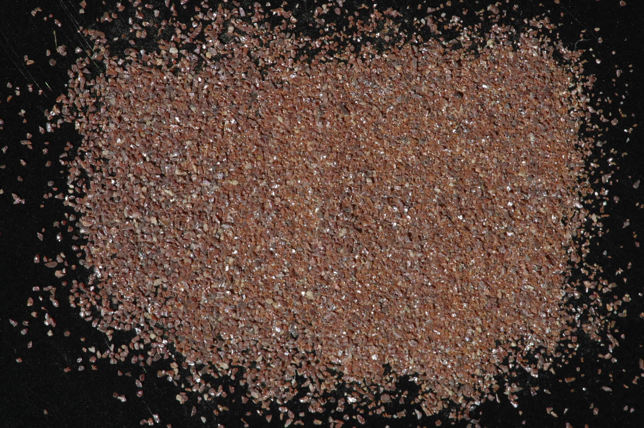
Sample photograph — USGS Spectral Library v7
Spectrum Details
- Modality
- NIR
- Category
- mineral
- Material Type
- Oxide
- Sample ID
- HS126.3B
- Collection Locality
- Oaxaca, Mexico
- Spectral Purity
- 1c2c3b4b # HS126.3B # 1= 0.2-3, 2= 1.5-6, 3= 6-25, 4= 20-150 microns
- Composition / XRD
- None # XRF, EPMA, ICP(Trace), WChem COMPOSITION_TRACE:
- Sample Description
- "0-15A. Rutile. Oaxaca, Mexico. (126B). Rutile, TiO2, is not a major rock-forming mineral, but is the commonest form of TiO2 in nature, and is very widely distributed as minute grains in many igneous and metamorphic rocks. Because of its resistance to weathering, it is also common in beach and river sands. TiO2 has a 3d° configuration, so that if a 3d band exists, it will normally be empty. It is possible to supply electrons to the 3d band by excitation from impurities, imperfections, or from the filled 2p band. The intense absorption in the blue is ascribed by Bevan and others (1958) as an exciton band arising from the transfer of an electron from an oxygen to a titanium. This sample contains 0.3% Fe, which does not appear to be located in a crystal field capable of producing the typical ferrous or ferric iron features near 1.0µ or 0.8µ. The steep fall-off in reflectivity toward the blue, which results in the reddish brown color common to rutile, is thus probably caused both by extrinsic absorption in the ferric ion and a sloping of the normal absorption edge in TiO2 because of defects and edge effects. Increasing amounts of ferric iron, niobium, and tantalum, which also enter the TiO2 lattice, result in deeper and deeper color, some varieties being almost opaque. The weak but sharp feature near 2.3µ must be due to some hydroxyl combination tone in this sample of rutile. It is an unusual band because no corresponding band near 1.4µ is apparent, and because chemical analysis of this sample yielded no measurable water. We conclude that a hydroxylated impurity is present in very small amount, and perhaps not at all in the portion of the sample analyzed." Hunt, G.R., J.W. Salisbury, and C.J. Lenhoff, 1971, Visible and near-infrared spectra of minerals and rocks: III. Oxides and hydroxides. Modern Geology, v. 2, p. 195-205. IMAGE_OF_SAMPLE:
- XRD Analysis
- Nearly pure sample of rutile with one or two trace impurities. One contaminant may be zirconium from grinding media. B. Benzel.
- X Units
- cm⁻¹
- Y Units
- Absorbance
- Data Points
- 2,126
Download
Attribution
USGS Spectral Library Version 7, U.S. Geological Survey
License: Public Domain
DOI: 10.3133/ds1035Analyze in Workbench
Open this spectrum in the SpectralBench workbench for advanced analysis, peak detection, and comparison.
Open Workbench