Siderite — FTIR1,801 points · 400–4000 cm⁻¹
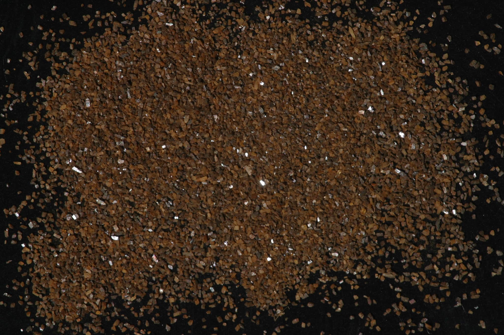
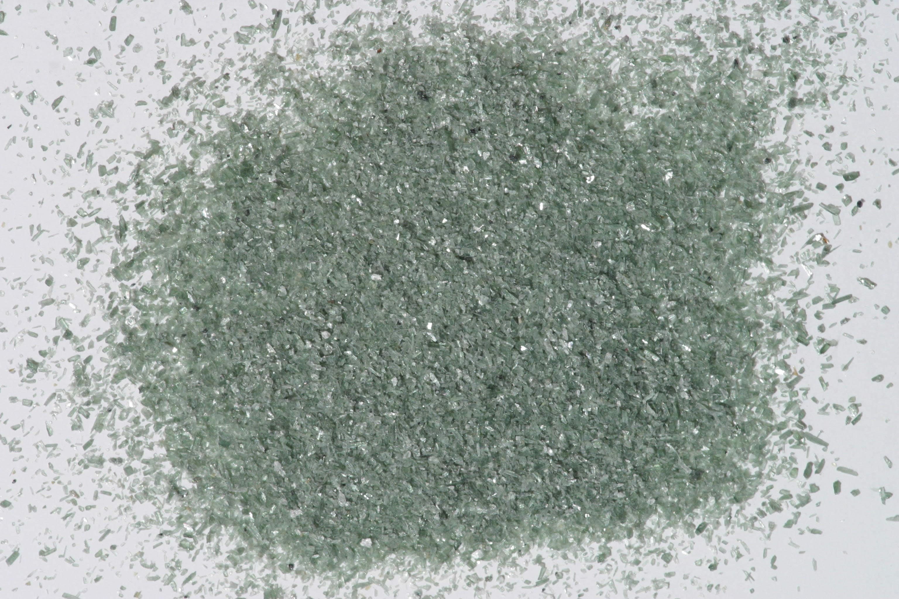
Sample photograph — USGS Spectral Library v7
Spectrum Details
- Modality
- FTIR
- Category
- mineral
- Material Type
- Carbonate
- Sample ID
- HS271.1B, HS271.2B, HS271.3B, HS271.4B
- Collection Locality
- Roxbury, Connecticut
- Spectral Purity
- 1a2_3_4_ # HS271.1B # 1= 0.2-3, 2= 1.5-6, 3= 6-25, 4= 20-150 microns SPECTRAL_PURITY: 1a2_3_4_ # HS271.2B # 1= 0.2-3, 2= 1.5-6, 3= 6-25, 4= 20-150 microns SPECTRAL_PURITY: 1a2a3a4_ # HS271.3B # 1= 0.2-3, 2= 1.5-6, 3= 6-25, 4= 20-150 microns SPECTRAL_PURITY: 1a2_3_4_ # HS271.4B # 1= 0.2-3, 2= 1.5-6, 3= 6-25, 4= 20-150 microns
- Composition / XRD
- None # XRF, EPMA, ICP(Trace), WChem COMPOSITION_TRACE:
- Sample Description
- Forms series with Magnesite and with Rhodochrosite. "C-7. Siderite. Roxbury, Conn. (68, hand-ground). Siderite, FeCO3, is found in extensive sedimentary beds, frequently contaminated with clay or organic matter. It is also commonly deposited in veins by hydrothermal solutions. Both magnesium and manganese commonly substitute for the iron. This particular sample is coarsely crystalline and medium brown in color. It is high in iron, as indicated by the strong ferrous ion band near 1.1µ. It is also slightly contaminated with magnetite. Being an opaque material, the magnetite lowers the overall reflectivity of the sample, makes the carbonate bands less prominent, and causes the crossover of the spectral curves in the visible. Analysis shows that this sample contains 0.98% Mg^2+ and 7.03% Mn^+2. However, the electronic transitions of iron dominate the spectrum to such an extent that none of the sharp features due to Mn^++ are apparent in the visible spectrum." NOTE: This a different sample number than this sample (271) but the collection locality is the same. Possibly a mislabeled sample. Hunt, G.R., J.W. Salisbury, 1971, Visible and near-infrared spectra of minerals and rocks: II. Carbonates. Modern Geology, v. 2, p. 23-30. Grain size fractions are indicated by the extension after the sample number: .1B = IMAGE_OF_SAMPLE:
- XRD Analysis
- Pure siderite Possibly some manganoan siderite also - patterns too similar to differentiate. Konnert, Judith and Marta Flohr, 1992, unpublished data, USGS Reston, VA.
- X Units
- cm⁻¹
- Y Units
- Absorbance
- Data Points
- 1,801
Download
Attribution
USGS Spectral Library Version 7, U.S. Geological Survey
License: Public Domain
DOI: 10.3133/ds1035Analyze in Workbench
Open this spectrum in the SpectralBench workbench for advanced analysis, peak detection, and comparison.
Open Workbench