Zoisite — FTIR1,801 points · 400–4000 cm⁻¹
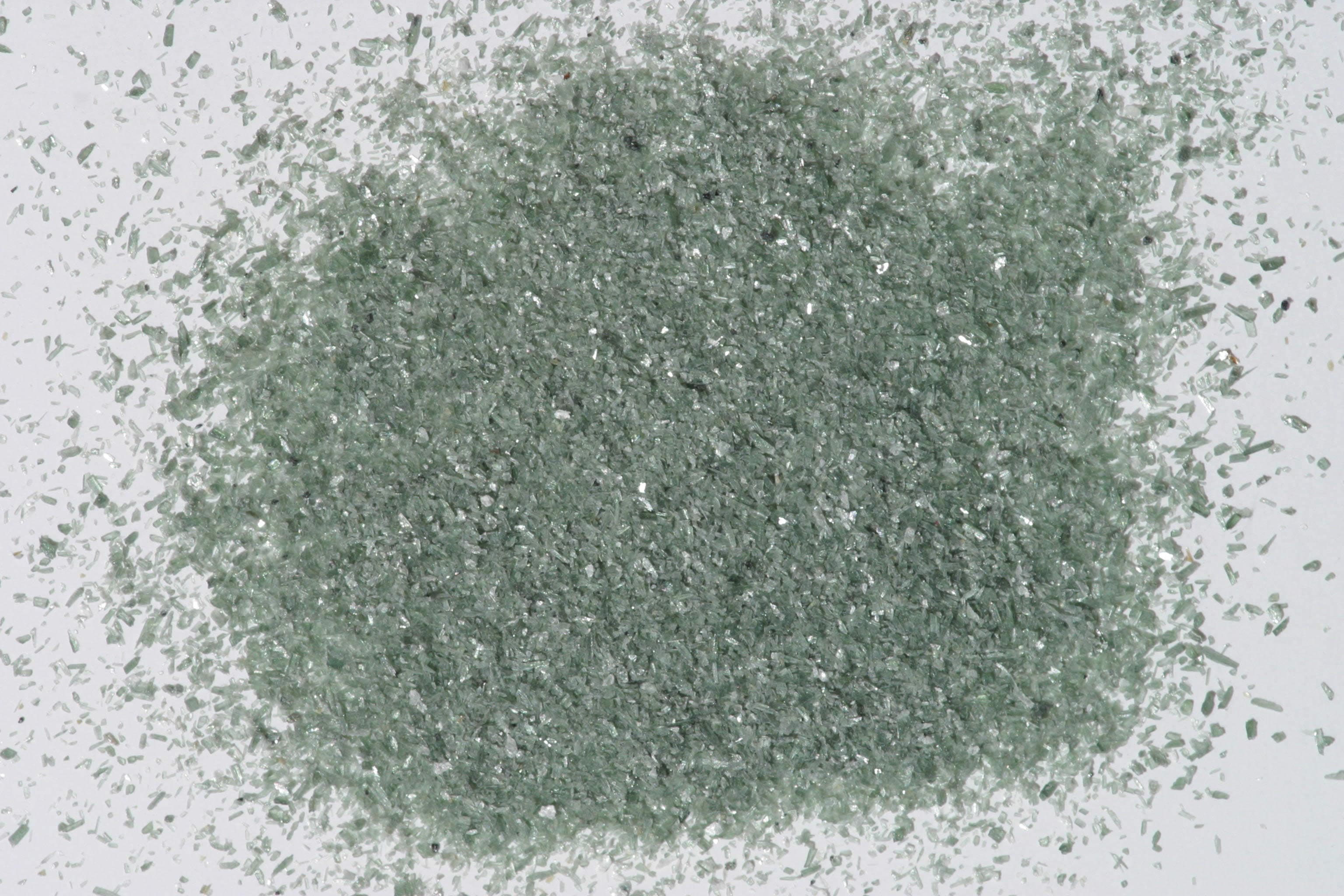
Sample photograph — USGS Spectral Library v7
Spectrum Details
- Modality
- FTIR
- Category
- mineral
- Material Type
- Sorosilicate
- Sample ID
- HS347.1B, HS347.2B, HS347.3B, HS347.4B, HS347.6
- Collection Locality
- Norway
- Spectral Purity
- 1b2_3_4_ # HS347.1B # 1= 0.2-3, 2= 1.5-6, 3= 6-25, 4= 20-150 microns SPECTRAL_PURITY: 1b2_3_4_ # HS347.2B # 1= 0.2-3, 2= 1.5-6, 3= 6-25, 4= 20-150 microns SPECTRAL_PURITY: 1b2b3c4c # HS347.3B # 1= 0.2-3, 2= 1.5-6, 3= 6-25, 4= 20-150 microns SPECTRAL_PURITY: 1b2_3_4_ # HS347.4B # 1= 0.2-3, 2= 1.5-6, 3= 6-25, 4= 20-150 microns SPECTRAL_PURITY: 1c2_3_4_ # HS347.6 # 1= 0.2-3, 2= 1.5-6, 3= 6-25, 4= 20-150 microns
- Composition / XRD
- None # XRF, EPMA, ICP(Trace), WChem COMPOSITION_TRACE:
- Sample Description
- "S-2 Zoisite 347--Norway. Ca2(AlOH)Al2-(SiO4)3. Zoisite, also a metamorphic mineral found in schists and gneisses, is orthorhombic and is dimorphous with clinozoisite. This sample displays extremely intense bands at 0.43, 0.53 and 0.8 µ, indicative of ferric iron possibly with contributions from Mn^+2. The major OH feature is most unusual, occurring at 1.68 µ, as opposed to the normal 1.4 µ position. There is also the unusual weak band at 1.85 µ, and the water feature at 1.9 µ with doubled features at 2.3 µ and 2.35 µ, and another band at 2.48 µ. These features are caused by combinations of OH with lattice or bending modes of ALOH, but the exact nature of these bands is not known." Hunt, G.R., J.W. Salisbury, and C.J. Lenhoff, 1973, Visible and near-infrared spectra of minerals and rocks: VI. Additional silicates: Modern Geology, v. 4, p. 85-106. Grain size fractions are indicated by the extension after the sample number: .1B = IMAGE_OF_SAMPLE:
- XRD Analysis
- Zoisite + medium amount quartz + small amount other(s); M: ~40% quartz, feldspar may be present
- X Units
- cm⁻¹
- Y Units
- Absorbance
- Data Points
- 1,801
Download
Attribution
USGS Spectral Library Version 7, U.S. Geological Survey
License: Public Domain
DOI: 10.3133/ds1035Analyze in Workbench
Open this spectrum in the SpectralBench workbench for advanced analysis, peak detection, and comparison.
Open Workbench